Tendon Diagram Simple : The Knee Joint Articulations Movements Injuries Teachmeanatomy - Vascular smooth muscle cells (vsmcs) are the stromal cells of the vascular wall and are responsible for regulating arterial tone, blood pressure, and blood supply of the tissues.
Tendon Diagram Simple : The Knee Joint Articulations Movements Injuries Teachmeanatomy - Vascular smooth muscle cells (vsmcs) are the stromal cells of the vascular wall and are responsible for regulating arterial tone, blood pressure, and blood supply of the tissues.. In this image, you will find frontalis, orbicularis oculi, zygomaticus, masseter, orbicularis oris, sternocleidomasteoid, deltoid, pectoralis major, biceps brachii, iliopsoas, adductor longus, gastrocnemius. The muscle stretch reflex is the most basic reflex pathway in the body and as such, understanding this allows understanding of more complex reflexes. When the muscles tighten (contract) they. The knee joint is a complex structure that involves bones, tendons, ligaments, muscles, and other structures for normal function. 1) tapping on the patellar.
Posted on may 16, 2016 by admin. The bones together make up the hip. A reflex is defined as an involuntary, unlearned, repeatable, automatic reaction to a specific stimulus which does not require input from the brain. Human body muscle system, the muscles of the human body that work the skeletal system, that are under voluntary control, and that are concerned with movement, posture, and balance. Attached to the bones of the skeletal system are about 700 named muscles that make up roughly half of a person's body weight.
Applied Basic Sciences Section 5 Postgraduate Orthopaedics from static.cambridge.org Tendons are the connective tissues between the bones and the muscles. The bones together make up the hip. The reflex arc governs the operation of reflexes. The muscles, tendons and ligaments. Diagram of artery with smooth muscle identification. The muscular system is responsible for the movement of the human body. Diagram of a chemical synapse between the reflex arc of the patellar or knee jerk reflex. Replicate the below figures with labels in your sketchbook.
Brings hip away from body.
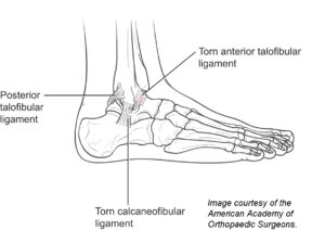
Ligaments attach one bone to another. Replicate the below figures with labels in your sketchbook. The foot diagram has a complex structure made up of bones, ligaments, muscles, and tendons.understanding the structure of the foot is best done by looking at a foot diagram where the anatomy has been labeled. Tendons in the knee play a very important role in holding the knee and the muscles together. Broadly considered, human muscle—like the muscles of all vertebrates—is often divided into striated muscle, smooth muscle, and cardiac muscle. When the muscles tighten (contract) they. Bones in shoulder, ligaments of the shoulder joint, parts of the shoulder joint, shoulder anatomy, shoulder joints and muscles, shoulder structure anatomy, shoulder tendon anatomy, shoulder tendons ligaments, human muscles, bones in shoulder, ligaments of the shoulder joint, parts of. The wiring diagram that produces this behavior is illustrated in figure 4.4.6. The muscular system is responsible for the movement of the human body. Extends spine and trunk back. When you finish with the above photo, head to the following model on posemanics and draw and label the linked model. The largest muscle masses in the leg are present in the thigh and the calf. Ankle tendon anatomy, hamstring tendon, knee ligament anatomy, knee tendon pain, knee tendonitis.
Ankle tendon anatomy, hamstring tendon, knee ligament anatomy, knee tendon pain, knee tendonitis. This diagram depicts muscle in the body 744×1054 with parts and labels. Lower body muscles diagram jpg 500 630 anatomiya cheloveka. When the muscles tighten (contract) they. The bones of the hip include the femur, the ilium, the ischium, and the pubis.
Foot And Ankle Muscle Strain Genesis Orthopedics And Sports Medicine from genesisortho.com Muscles tendons and ligaments run along the surfaces of the feet allowing the complex movements needed for motion and balance. The muscles at the top of the foot fan out to supply the individual toes. If you would like to learn all the parts of the foot structure, you have come to the right place. Posted in diagrams, muscles | tagged human muscles, human muscles anatomy, muscle, muscle chart, muscle diagram, muscles, muscles anatomy, muscles diagram, muscles system anatomy female 1024×1111. The tendons are thick bands that connect muscles to bones. 17 photos of the diagram of shoulder muscles and tendons. Raises and rotates arm in all directions. Structure and function of ligaments and tendons.
Posted on may 16, 2016 by admin.
Tendons are the connective tissues between the bones and the muscles. Attached to the bones of the skeletal system are about 700 named muscles that make up roughly half of a person's body weight. The muscle stretch reflex is the most basic reflex pathway in the body and as such, understanding this allows understanding of more complex reflexes. Related posts of simple human muscle diagram. Ligaments and tendons are both made of connective tissue and both can be torn or overstretched, but they differ in function. Brings hip away from body. Superficial and deep anterior muscles of upper body superficial and deep posterior muscles of upper body. Muscles of the hip anatomy pictures and information. The muscles, tendons and ligaments. A tendon is a band of tissue that connects a the two. Anatomy of the hamstring upper leg. Tendon, tissue that attaches a muscle to other body parts, usually bones. When there is damage to one of the structures that surround the knee joint, this can lead to discomfort and disability.
The muscles are located mainly in the sole of the foot and divided into a central (medial) group and a group on either side (lateral). It attaches to the wrist bone, the pisiform, and as well as the 5th hand bone. Nervous system 3, reflex arc. Tendon, tissue that attaches a muscle to other body parts, usually bones. A simple model illustrating the dependence of ligament/tendon nonlinear stress/strain relationships is shown below:
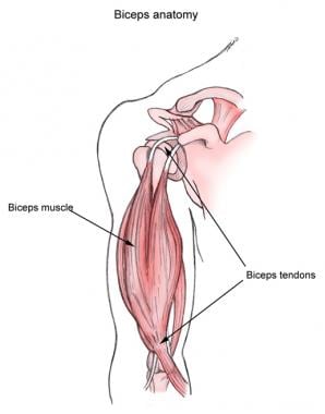
Bicipital Tendonitis Background Epidemiology Functional Anatomy from img.medscapestatic.com Flexes elbow and moves forearm. Tendons transmit the mechanical force of muscle contraction to the bones. Tears of the achilles tendon can be tiny (microtears), or large, causing pain, swelling, and impaired movement. The foot diagram has a complex structure made up of bones, ligaments, muscles, and tendons.understanding the structure of the foot is best done by looking at a foot diagram where the anatomy has been labeled. In this case, as a spring is stretched to its limit its stiffness increases. Ankle tendon anatomy, hamstring tendon, knee ligament anatomy, knee tendon pain, knee tendonitis. In this image, you will find frontalis, orbicularis oculi, zygomaticus, masseter, orbicularis oris, sternocleidomasteoid, deltoid, pectoralis major, biceps brachii, iliopsoas, adductor longus, gastrocnemius. Each of these muscles is a discrete organ constructed of skeletal muscle tissue, blood vessels, tendons, and nerves.
Brings hip away from body.
The tendons are thick bands that connect muscles to bones. Related online courses on physioplus. Raises heal when leg is straight. Posted on april 3, 2019april 3, 2019. Ligaments attach one bone to another. This diagram depicts muscle in the body 744×1054 with parts and labels. 1) tapping on the patellar. The foot diagram has a complex structure made up of bones, ligaments, muscles, and tendons.understanding the structure of the foot is best done by looking at a foot diagram where the anatomy has been labeled. *click them to make them larger & view details. Tendinitis is inflammation or irritation of a tendon — the thick fibrous cords that attach muscle to bone. A simple model illustrating the dependence of ligament/tendon nonlinear stress/strain relationships is shown below: Tendons transmit the mechanical force of muscle contraction to the bones. Human muscle system, the muscles of the human body that work the skeletal system, that are under voluntary control, and that are concerned with movement, posture, and balance.


0 Komentar